Catalog # SP1641
Sphingosine Kinase 1 (Ser-225), phospho-specific Antibody
Rabbit Polyclonal
| ELISA | 1:2000 |
| WB | 1:1000 |
Size 100 μl
Species Reactivity Hu
MW 40-50 kDa
Sphingolipids are metabolized into bioactive products that include ceramide, sphingosine, and sphingosine-1-phosphate (S1P). Sphingosine Kinase (SK) catalyzes the phosphorylation of the lipid sphingosine, creating S1P. S1P subsequently signals through cell surface G protein-coupled receptors, as well as intracellularly, to modulate cell proliferation, survival, motility and differentiation. Two isoforms of SK have been identified, SK1 and SK2. The mRNA for both of these isoforms is widely expressed with SK1 expression highest in brain, heart, kidney, thymus, spleen and lung, while SK2 is highest in kidney and liver. SKs can be activated through growth factor, G protein-coupled, and immunoglobulin receptor signalling. SK1 has been shown to mediate cell growth, prevention of apoptosis, and cellular transformation, and is upregulated in a variety of human tumors. Regulation of SK1 may occur through ERK mediated phosphorylation of Ser-225. This phosphorylation leads to increased activity and translocation to the plasma membrane.
References
Pitson, S.M. et al. (2005) J Exp. Med. 201(1) :49.
Pitson, S.M. et al. (2003) EMBOJ. 22(20) :5491.
Melendez, A.J. et al. (2000) Gene 251(1) :19.
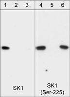
Western blot image of recombinant his-tagged human SK1 protein that was phosphorylated with ERK-2. Blots were probed with anti-SK1 (Central Region) (SP1621; lanes 1-3) and anti-SK1 (Ser-225) (SP1641; lanes 4-6). Both antibodies were used in the presence of no peptide (lanes 1 & 4), phospho-SK1 (Ser-225) peptide (SX1645; lanes 2 & 5), or unphosphorylated SK1 (Ser-225) peptide (SX1625; lanes 3 & 6).

Western blot of HeLa stimulated with calyculin A (lanes 1-4). The blots were untreated (lane 1 & 3) or treated with lambda phosphatase (lane 2 & 4), then probed with anti-SK1 (Central region) SP1621 (lanes 1 & 2) or anti-SK1 (Ser-225) SP1641 (lanes 3 & 4).
*For more information, see UniProt Accession Q9NYA1
The products are are safely shipped at ambient temperature for both domestic and international shipments. Each product is guaranteed to match the specifications as indicated on the corresponding technical data sheet. Please store at -20C upon arrival for long term storage.
*All molecular weights (MW) are confirmed by comparison to Bio-Rad Rainbow Markers and to western blot mobilities of known proteins with similar MW.
Product References:
Yamada, A. et al. (2018) Mol Cancer Res. 16(6):1059. (ICC/IHC: human breast tumors)Fu, P. et al. (2016) J Biol Chem. 291(53):27187. (WB/ICC: human endothelial cells)
Durham, J. et al. (2015) Invest Ophthal Vis Sci. 56(6):3441 (WB: human retinal cells and pericytes)
Wilson, P.C. et al. (2015) Mol Endocrinol. 29(6):896 (WB: rat aortic vascular smooth muscle cells)
Sun, K. et al. (2015)Blood 125(10): 1643. (WB: mouse erythrocyte)
Xiu, L. et al. (2015) Am J Pathol. 185(2):387 (WB: hMSC cells)
Kim, E.Y. et al. (2014) FASEB J. 28(10): 4347 (WB: HEK293-CD40 cells)
Dai, L. et al. (2013) Molec Endocrinology 28(2): 197. (WB: human MCF7 breast cancer)
Seo, Y. et al. (2013) PLoS ONE 8(8):e75005. (WB: MDCK, A549, and HEK293)
Yasuo, M. et al. (2013) PLoS One 8(1):e53927. (WB: rat lung)
Park, K. et al. (2013) Mol Cell Biol. 33(4)752. (WB: human keratinocytes)
El-Shewy, M. et al. (2012) Mol Endocrinol 26(5):833-45. (WB: rat glomerular mesangial cells)
Mair, K. et al. (2010) British J Pharmacol 161:176. (WB: rat coronary artery)
Weis, T. et al. (2010) European J Cell Biol 89(10):733. (WB: HUVECs )
Benakanakere, M. et al. (2010) PLoS ONE 5(7):e11512. (WB: human Keratinocytes )
Alvarez, S.E. et al. (2010) Nature 465:1084. (WB: A7 melanoma cells)
Hengst, J. et al. (2010) Archives Biochem Biophys 494(1):23. (WB: SK1 transfectants: )
Shida, D. et al. (2008) Cancer Res. 68(16):6569. (WB: human MNK1 cells + LPA)
Miller et al. (2008) Mol Cell Biol 28(12):4142. (WB: human esophageal carcinoma)
Sobue, S. et al. (2008) International J Hematol 87(3):266. (WB: leukemia cell lines: )
Paugh, B.S. et al. (2008) FASEB J. 22(2):455. (WB: human glioblastoma A172)
This kit contains: