Catalog # TM4111
α-Tubulin (C-terminus) Antibody
Mouse Monoclonal
| ELISA | 1:2000 |
| ICC | 1:200 |
| IHC | 1:200 |
| IP | 1:100 |
| WB | 1:1000 |
Size 100 μl
Species Reactivity Hu, Rt, Ms
MW 50 kDa
Isotype IgG1
Microtubules (MTs) are cytoskeletal elements that play an essential role in cell division and cytoplasmic organization. MTs are dynamic polymers of a/β-Tubulin heterodimers. At least two populations of MTs, called dynamic and stable according to their rates of turnover, are readily distinguishable in cells. The proteins associated with MTs (MAPs) are among the best-known factors that regulate MT dynamics and stability. In addition, a variety of different post-translational modifications may also regulate MT dynamics and stability. Phosphorylation is one of these modifications and it can occur on serine, threonine, and tyrosine residues in α- and β-Tubulin isoforms. Multiple kinases can phosphorylate Ser-444 at the C-terminus of βIII-Tubulin in vitro, and unphosphorylated Ser-444 may be an early marker for cells of neuronal lineage. Cdk1 can phosphorylate Ser-172 in β-Tubulin during mitosis and this may impair tubulin incorporation into microtubules. In α-tubulin, PKC can phosphorylate Ser-165 leading to increased cell motility in human breast cells.
References
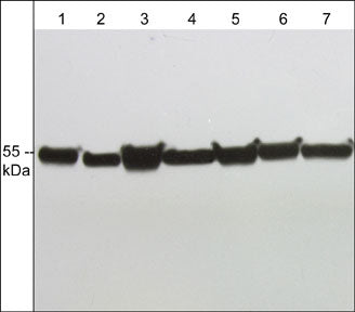
Western blot analysis of α-tubulin expression in human A431 (lane 1), HUVEC (lane 2), Jurkat (lane 3), mouse J774.1 (lane 4), human PC-3 (lane 5), rat PC12 (lane 6), and mouse C2C12 (lane 7). The blot was probed with anti-α-Tubulin (C-terminus) at 1:1000.

Immunocytochemical labeling of α- and βI-Tubulin in rat A7r5 cells. The cells were labeled with anti-βI-Tubulin (TM1541) (left) and anti-α-tubulin (TM4111) (right). The antibodies were detected using Goat anti-Mouse conjugated to DyLight® 488.
*For more information, see UniProt Accession Q9BQE3
The products are are safely shipped at ambient temperature for both domestic and international shipments. Each product is guaranteed to match the specifications as indicated on the corresponding technical data sheet. Please store at -20C upon arrival for long term storage.
*All molecular weights (MW) are confirmed by comparison to Bio-Rad Rainbow Markers and to western blot mobilities of known proteins with similar MW.
This kit contains: